Foot and Ankle pain
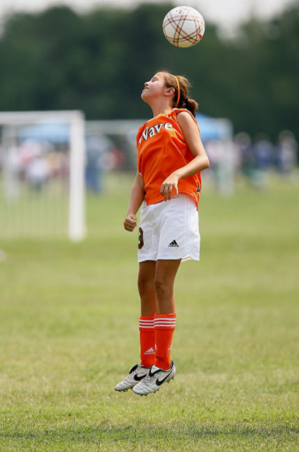
Children and adolescents’ bodies are forever changing. As children grow strength, flexibility, coordination and skill can fluctuate and thus it puts them at risk for injury and pain.
We are keen to get kids back in the game as quickly as possible so the appropriate education, load management and treatment is often tailored to you son or daughter. Discussing a return to activity plan with your child’s PE teacher or sporting coach will form part of this plan.
Common conditions we treat.
- Stress fractures due to overuse
- Sprained ankles
- Growing pains
- Tarsal coalition (where two bones are joined together across a joint)
- Pain at the heel or side of the foot with activity is generally caused by apophyseal injuries such as Sever’s disease or Iselin’s disease
- Pain at the top of the foot or under the ball of the foot that is caused by avascular necrosis. These conditions are called Kohler’s disease and Freiberg’s disease
- Accessory bones (additional ossicles of bone in spots that can cause pain)
- Inflammatory joint pain
- Post-surgical pain
Below are two common reasons children present with pain without injury.
Calcaneal Apophysitis (Sever’s Disease).
Within the lower limb are a number of open growth plates and often children aged 8-15 can have pain at these spots. The two most common tibial tuberosity apophysitis (Osgood Slatters disease) and calcaneal apophysitis (Sever’s Disease). Generally, this cause of pain are a self-limiting condition that children and young adolescents grow out of while allied health clinicians can help manage the symptoms.
The symptoms that kids experience is pain at the heel with, and shortly after activity that goes away with rest. Children often present with symptoms of calcaneal apophysitis at the start of the school year, when there is cross over from winter to summer sporting codes, a rapid increase in activity or after a period of minimal activity.
Often the change in footwear type can be an external contributor; football boots, soccer boots and basketball shoes most commonly as these tend to be very flat in design. Or as a child starts an activity or sport that they have not done before and are building their endurance for that activity.
Although much of the literature will say that calcaneal apophysitis affects very active children, in my experience I have seen it in both very active and junior elite sporting children and those that are requiring a lot of motivation to participate and move around.
There is no set algorithm or treatment flow chart for this condition although there are some common first steps parents can try; ice pack after activity, gel heel cups from the pharmacy or sports store and if there has been a rapid increase in activity consider modify a session or two for a week. Parents should be aware night pain, swelling, heat or limping will need to be seen by a health professional as this may not be calcaneal apophysitis.
Once we have a confirmed diagnosis, we address the other factors that may be contributing such as training and activity loads, strength, flexibility, footwear type, foot posture and running style. I would set up a treatment plan that may include a strength and flexibility program, modification of activity or progression back to normal activity, unload the heel with a heel raise or heel cup, orthotics or a change in footwear.
Accessory Bones
Within the foot there are a number of spots to have “accessory” ossicles. These are a normal variant that are often misdiagnosed as a fracture.
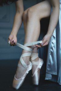
The most common in the foot is the Os Trigonum that can be seen in up to 25% of people. This accessory ossicle sits at the back of the ankle can often cause irritation to the surrounding soft tissue and joint structures after a nasty ankle injury or in sports where going up onto your toes a lot such has ballet, netball, martial arts.
The accessory that I see most in the clinic and causes the most discomfort in my patients is the Os Tibiale. This accessory ossicle can present in three types.
- Type one is where the additional bone sits just off the navicular bone within a tendon and is usually symptomatic
- Type two is where the additional bone sits just off the navicular bone, but it connected to navicular by cartilage or fibros tissue. Often one of the main tendons that holds up the arch of the foot inserts into the ossicle. This can cause pain as the ossicle is pulled away from the main bone by the tendon
- Type three is where the additional bone appears more line a enlarged navicular or a fused type 2. This can cause irritation and pain.
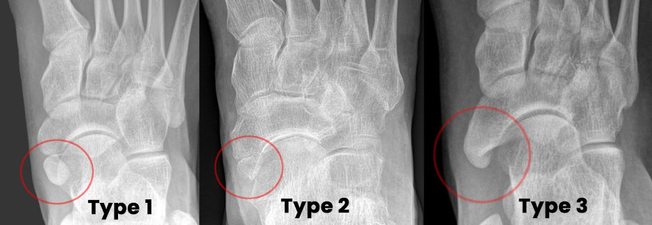
By Hellerhoff – Own work, CC BY-SA 3.0, https://commons.wikimedia.org/w/index.php?curid=39354099
Both these types of accessory ossicles are often treated by addressing the factors that may be contributing to the pain such as training and activity loads, strength, flexibility, footwear type, foot posture and running style. I would set up a treatment plan that may include a strength and flexibility program, modification of activity or progression back to normal activity, unload the ossicle with an orthotic or a change in footwear.